

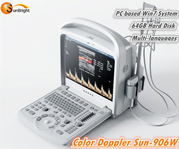
Full imaging mode:CFM, PDI, PW, HPRF, etc. Standard two transducer sockets with 15aa LCD monitor iBeamTM,iZoomTM
Features
l Full imaging mode 2d, CFM, PDI, PW, HPRF, etc.
l Standard two transducer socket with 15' LCD monitor
l Full function control panel with black-lit and 8 level TGC
l Tissue Harmonic Imaging
l iBeamTM spatial compounding imaging reduces random noise and enhances border differentiation without compromising frame rate
l iZoomTM automatically expands the image to full screen
l Full patient database solutions: DICOM3.0, AVI/JPG, USB2.0, HDD, PDF report, etc.
Specification:
l Display 15" LCD monitor
l Display model B, 2B, B/M, 4B, B/C
l TGC: 8-section TGC adjustment
l Scanning depth 24mm
l Image processing preset, PRF, gain, Dynamic range, gray map, Black/white, Left/right, Up/down
l Digital technology DRF&DFS
l Transducer frequency 2.5-10MHz
l Gray scale 256
l Cine-loop 100 frame cine loop memory
l iTouch One key for optimization
l iClear One key for clearer
l Zoom Pan zoom
l Probe connector 3 in all, 1 for update in future
l USB ports 4
l Power supply 100-240VAC± 10%, 50Hz/60Hz
Standard Configurations:
l Main unit
l 15"LCD with high resolution and contrast
l 64GB hard disk
l 2GB DDR Ram
l INTER Core 2Duo L9400CPU
l 4 USB ports, 1 S-video output port
l 2 probe connectors for use, one for update
l Measurement and Calculation software package ( General, OB/GYN. small parts, Urology)
l 3.5MHz Electronic convex array probe
Probe Types:
R50 3.5MHz broadband abdominal convex probe
Applicable to: examination and diagnosis of abdominal tissue including liver, gallbladder, spleen, pancreas, kidney, gastrointestinal tract, uterus, bladder, prostate and other organs, as well as the measurement of fetal growth and maturity in obstetric diagnosis.
R20 3.5MHz broadband cardiac micro-convex probe
Applicable to: cardiac, peritoneal tissues and structure.
R10 6.5MHz broadband convex endoprobe
Applicable to: Vagina and rectum, such as uterus, rectum, rectum wall and prostate.
L40 7.5MHz broadband linear probe
Applicable to: neonate, muscular skeletal system, peripheral blood vessel and organella (including uber,thyroid, testis, etc.).
Measurement and calculation items:
Abdomen: LIVER , GB gall bladder, GBWT gall bladder wall thickness, CBD common bile duct, PORTAL VEIN , SPLEEN, RT kidney, LT kidney
Obstetrics measurement:
1.Selection of fixed database version
2.Edit of customized database
3.Calculation of gestational age and estimation of expected date of childbirth
4.Calculation of fetus weight
5.Infer the gestational weeks and expected date of childbirth according to LMP
6.Infer the gestational weeks and expected date of childbirth according to BBT
7.Fetal biophysical score
8.Fetus growth curve
Gynaecology measurement(pevis menu)
Mainly to measure uterus. Measure its cervix length, endometrium thickness, uterus length (UTL), uterus width(UTW), uterus height(UTH). Meanwhile, it can measure left/right ovary and cyst.
Multiple left/right follicle measurement
Provides an effective method to detect follicle developmental abnormality and ovulation, give comparatively definite diagnosis on follicle development abnormality and various ovulatory disorders.
Pelvis measurement
Skeletalmuscle measurement
Skeletalmuscle is also called as striated muscle, as one kind of muscle.
Urology measurement
lithangiuria, tumor, deformity, infection, trauma, kidney transplant, male reproduction etc..Urology internal medicine is mainly responsible for diagnosis and treatment of kidney disease, kidney, nephritis, rental function incomplete, uremia etc..
Peripheral blood vessel measurement
Refers to trunk and arms/legs blood vessels except cardiovascular and cerebric blood vessel.
Small parts measurement
Small parts measurement includes "Thyroid, testicle" volume measurement , "Isthmus" measurement.
Cardiac measurement
Conventional AORTA (AO): Heart (PROX AO AP) Left Ventricular Posterior Wall (AO) Apex Cordials (AP)