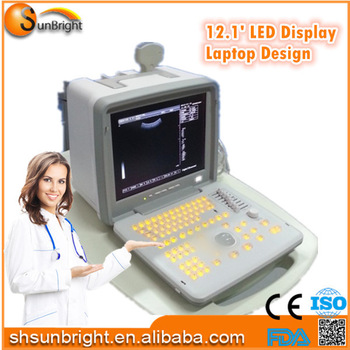


Specification:
|
Display modes |
B, B/B, 4B, B+M, M |
|
Image multiplying factor |
3.5MHz/R60 convex array probe: ×0.8, ×1.0, ×1.2, × 1.5, ×1 .8, ×2.0 (six modes) ×0.8, ×2.0 (display penetration depth); 6.5MHz/R13 Dimpling array probe: ×0.8, ×1.0, ×1.2, × 1.5 (four modes); 7.5MHz/L40 Line array probe: ×0.8, ×1.0, ×1.2, × 1.5 (four modes). |
|
E-zoom |
magnify 2 times of real time image |
|
Dynamic range |
0~120dB adjustable |
|
Focus position |
1, 2, 3 and 4-segments dynamic electron focusing |
|
Image processing |
γ correction, frame correlation, point correlation, line correlation, digital filtering, digital edge enhancement and pseudo color processing, etc. |
|
Frequency conversion |
2.5MHz/3.0MHz/3.5MHz/4.0MHz/5.0MHz five periods of frequency conversion; frequency range applies 5.5 MHz/6.0 MHz/6.5 MHz/7.0 MHz/7.5MHz can match high frequency probe. |
|
Measuring function |
Distance, circumference/area (method of ellipse, method of loci), volume, heart rate, gestational weeks (BPD, GS, CRL, FL, AC), expected date of confinement and fetus weight, etc. |
|
Annotation function |
hospital name, patient’s name, age and gender; 64 body marks (with probe’s position); Full-screen character annotation; Real-time clock display; |
|
Puncture guide |
3.5MHz convex array probe can display puncture guide line in B mode. |
|
Gain control |
8 segments TGC and overall gain can be adjusted respectively. |
|
Image polarity |
left and right flip, black and white flip, up and down flip. |
|
Capacity cine loop |
real time display 256 consecutive images which are memorized successively |
|
Image playback |
series playback or check one by one |
|
Permanent storage |
127 images |
|
Output interface |
SVGA video output offers connection to SVGA color display, PAL video output offers connection to PAL display, video printer, video image recorder and image workstation, etc |
|
USB port |
offers storing images in USB flash disk |